

Vena8 Fluoro+
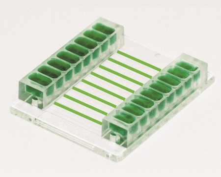
The Vena8 Fluoro+™ biochip is ideal for rolling and adhesion assays on protein coatings or cell monolayers. Ideal for thrombosis; whole blood assays. Compatible with immunofluorescence and confocal microscopy.
Features:
• Disposable plastic biochips for cell analysis of cells under shear flow including rolling and adhesion assays on protein coatings or cell monolayers.
• Immunofluorescence staining of living and fixed cells.
• Ideal for whole blood, primary cells, rare cells e.g. where samples are more difficult to retrieve or culture or only small sample volumes available.
• Perfect for high flow rates / high shear stresses e.g. platelet adhesion, aggregation and thrombi formation studies for thrombosis using whole blood. In standard flow chambers / perfusion chambers; significant volumes of blood would be required making the assay impossible.
• No assembly of the biochip is required unlike many standard perfusion chambers / flow chambers.
• No luer lock connections which increase dead volume. Cellix’s biochips have a unique plug & play connection with tubing connections which are autoclaveable and reuseable.
Applications: Flow assays using minimal sample volumes (whole blood, cell suspensions, proteins etc.); immunofluorescence and confocal microscopy.
Specifications:
| Substrate thickness | 0.17mm = 170μm |
| Substrate material | High quality plastic, compatible with immunofluorescence and confocal microscopy |
| Number of channels per biochip | 8 |
| Height of channels | 0.1mm = 100μm |
| Width of channels | 0.4mm = 400μm |
| Length of channels | 28mm = 2.8cm |
| Volume of each channel | 0.8μL |
| Volume of protein required for coating | 10μL |
| Volume of reservoir to hold sample | 0.1mL; also possible to connect via tubing to eppendorf if greater sample volume or high shear stress / flow rate assay is being performed |
Vena8 Fluoro+ Biochip Tutorial
Vena8 Fluoro+ biochip protocol, ideal for thrombosis assays, Vena8 Fluoro+ biochip is compatible with confocal microscopy and immunostaining. Tutorial illustrates coating of biochip with adhesion molecules (e.g. VWF, collagen, fibrinogen, VCAM, ICAM etc.); conducting an assay with cell suspension (e.g. platelets, whole blood, T-cell, monocytes, neutrophils, eosinophils, PBMCs etc.) and analysis of data. Quantitate data outputs include IC50 curves of cell adhesion (e.g. platelet adhesion, aggregation, thrombi formation) vs. shear flow rate (mimicking in vivo arterial/venous/capillary flow); morphology analysis of cell adhesion and migration; tracking of flowing, rolling and migrating cells. Ideal for functional cell-based assays for cell-ligand interactions; preclniical drug development and lead optimization. *** NO AUDIO ***