Afyon SDS-PAGE Örnek Hazırlama Kiti
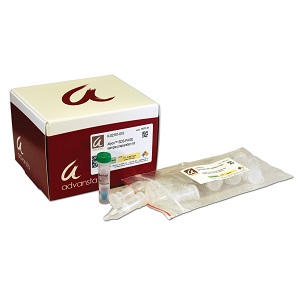
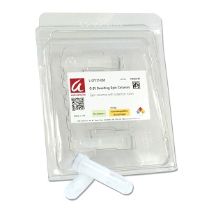
Kit for fast, easy concentration and purification of samples for SDS-PAGE
• Purify – Remove contaminants that can interfere with electrophoresis (GuHCl, urea, Ammonium sulfate, etc)
• Quick – Protein samples ready to load in less than 10 minutes
• Safe – and non-toxic – no DMSO required
• Compatible with SDS-PAGE and downstream Western blotting
• Many times faster than concentration and buffer exchange via ultrafiltration or dialysis
Description
Concentrate and purify protein samples for SDS-PAGE with the Afyon sample purification kit
The Afyon SDS-PAGE sample preparation kit provides a means to quickly concentrate protein samples, and separate them from buffers that interfere with electrophoresis. The fast, efficient protocol generates samples ready to load on a gel in less than ten minutes (Figure 1), much more quickly than can be achieved with alternate methods such as dialysis, acetone or TCA precipitation. Additionally, the Afyon protocol is easily scaled up, allowing multiple samples to be prepared in parallel. Samples remain compatible with both chemiluminescent and fluorescent Western blotting (Figure 2). Purification successfully removes comtaminants that can interfere with electrophoresis (Figure 3) an shows similar results to ultracentrifugation while being ten times faster (Figure 4).
Prepare samples in as little as 10 minutes.
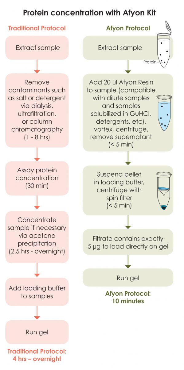
Figure 1: Afyon protocol compared to traditional purification protocol.
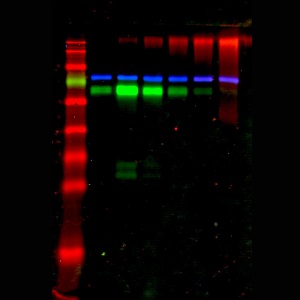
Afyon concentration is compatible with Western blot detection.
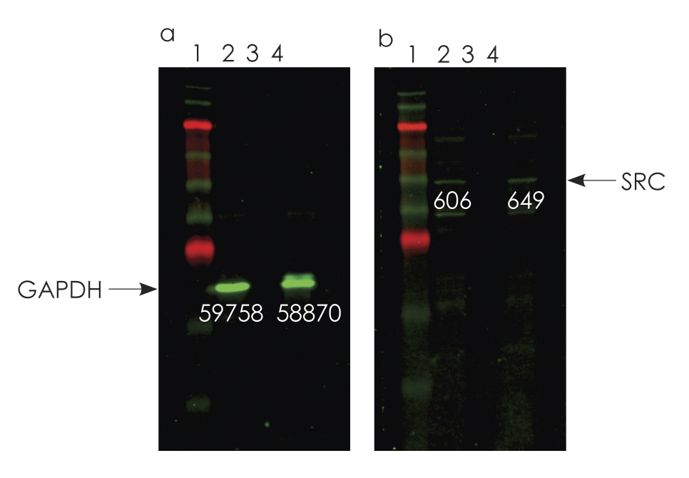
Figure 2: Western blots were created with samples of A431 cell extract (panels a and b, lane 2), diluted cell extract (lane 3) and the diluted extract concentrated using the Afyon protocol (lane 4). The blots were detected using the WesternBright MCF multicolor fluorescent Western blotting kit. No bands were detectable in the diluted cell extract when stained for either GAPDH (panel a, lane 3) or SRC (panel b, lane 3). However, after Afyon concentration, bands are easily visualized for both proteins (lanes 4). Band intensities are indicated.
Afyon removes contaminants that can interfere with electrophoresis.
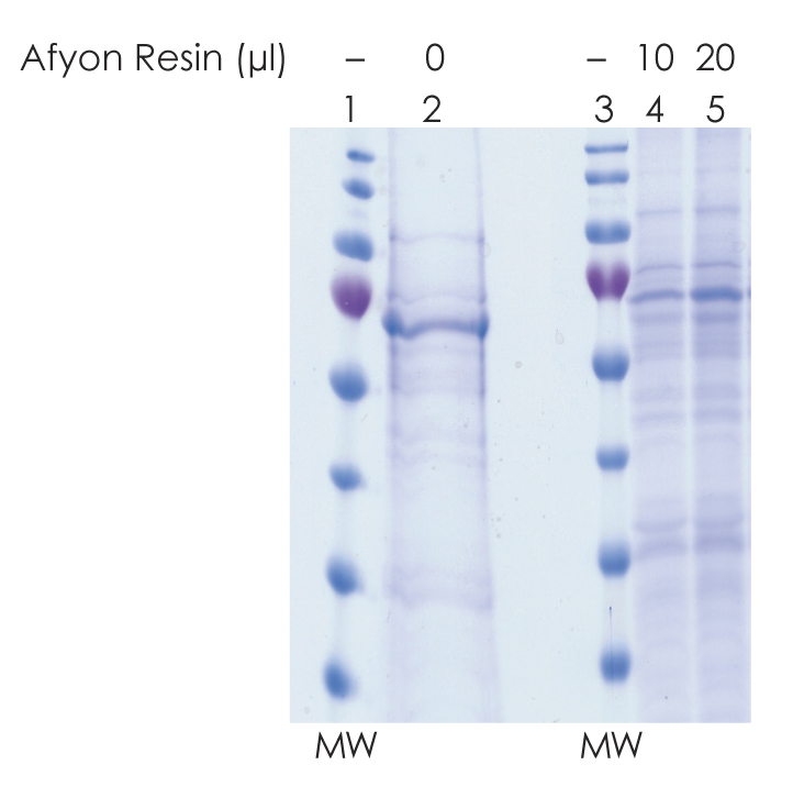
Figure 3:Afyon removes buffer components such as guanidinium chloride, thiocyanate or urea that can cause samples to migrate irregularly. A sample of K-561 cell lysate in 4 M guanidinium thiocyanate spreads and runs irregularly on a gel (lane 2). When the protein is recovered using Afyon resin (lanes 4-5), the salt is removed and the sample runs cleanly. 10 μl of Afyon recovers 2.5 μg protein (lane 4), while 20 μl of Afyon recovers 5 μg protein (lane 5). MW = molecular weight markers.
Concentrate samples 10 times faster than spin ultrafiltration.

Figure 4:. HeLa cell lysate was diluted to 20 μg/ml with 1M NaCL, 20 mM Tris pH 7.6. 1 ml diluted lysate was concentrated and buffer exchanged using an ultrafiltration spin filter (Milipore), or Afyon resin. Ultrafiltration took over one hour, while Afyon took less than 10 minutes. Lane 1: molecular weight markers. Lane 2: sample concentrated by ultrafiltration. Lane 3: sample concentrated using Afyon.


