

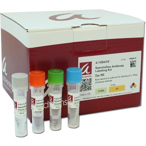
SpectraDye Antikor İşaretleme Kiti
Make your own fluorescent antibody in one easy step.
• Quick and easy – one-step labeling in 30 minutes
• Versatile – Labeled antibodies are compatible with immunofluorescence microscopy, flow cytometry, Western blotting
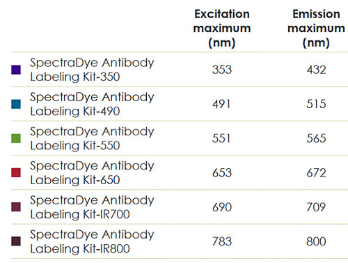
• Flexible – choose from 6 commonly used fluorescent dyes
• Save time and money – no secondary or extra steps, once you label your primary
• Save antibody – labeling reaction requires as little as 10 µg antibody
Kit includes:
Antibody Labeling Buffer
SpectraDye Dye Solution*
Quenching Solution
Neutralization Buffer
Sufficient reagents to label up to 1 mg of antibody
*Except kit K-11060-010
Description
SpectraDye Antibody Labeling Kits include sufficient reagents to label up to 1 mg of antibody. SpectraDye dye kits contain Antibody Labeling Buffer, SpectraDye Dye Solution, Quenching Solution, and Neutralization Buffer. The user-supplied dye kit does not contain dye, but does contain high-quality DMSO to prepare your own dye solution.
What is the SpectraDye Antibody Labeling kit useful for?
Lower background in immunofluorescence-based experiments: Using a secondary antibody can result in high non-specific binding and low reproducibility. Switching to a directly labeled primary antibody can improve your results.
Single-color fluorescent applications: Direct labeling of your antibody increases reproducibility, and allows the same antibody to be used in various applications to validate results.
Multi-color fluorescent applications: With 6 dyes to choose from, SpectraDye Antibody Labeling Kits give you tremendous flexibility, and since you don’t need secondary antibodies, the species of your primary antibody does not matter.
What does the labeling reaction involve?
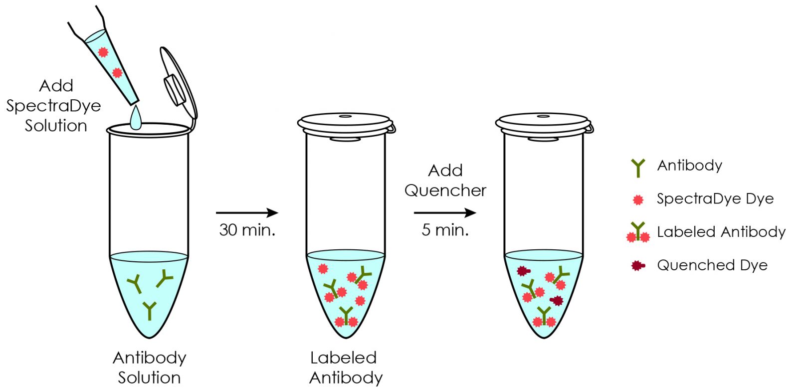
The labeling reaction really is fast and easy. Start by diluting antibody in PBS to 1 mg/ml. Next, add 25 μl Antibody Labeling Buffer to 100 μl antibody solution. Mix thoroughly by pipetting up and down. Add 12 μl of Dye Solution to the reaction and mix by pipetting. Incubate 30 min at room temperature. Additional recommended steps: Add 3 μl Quenching Solution to the reaction mixture and incubate for 5 min at room temperature; then, add 8μl Neutralization Buffer to the reaction mixture. Antibody is labeled and ready to use.
Experimental applications:
Immunofluorescence Microscopy: Decrease non-specific binding and eliminate species cross-reactivity when analyzing multiple antigens on the same slide.
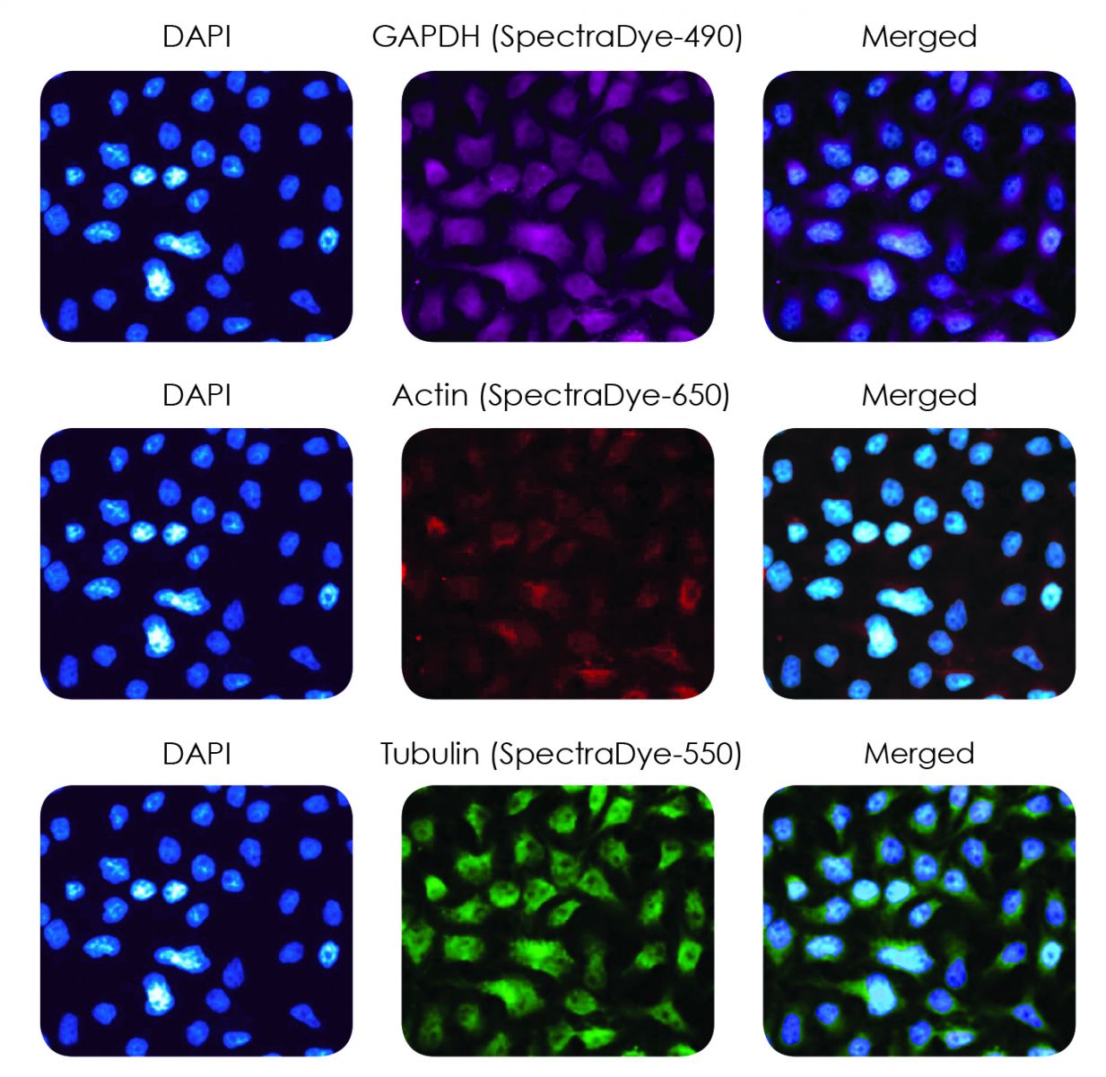
Increase the specificity of multicolor stained slides by using high quality monoclonal antibodies. HeLa cells were fixed on a glass coverslip then blocked with neutral donkey serum. The cells were then stained simultaneously with a panel of high-affinity SpectraDye labeled mouse monoclonal antibodies. The anti-GAPDH antibody was labeled with SpectraDye-490 (magenta), the anti-actin antibody was labeled with SpectraDye-650 (red) and the anti-tubulin antibody was labeled with SpectraDye-550 (green). The nuclei were stained with DAPI (blue).
Flow Cytometry: Simplify, decrease variability, and improve data quality in your flow cytometry assays by using your own fluorescently-labeled primary antibodies.
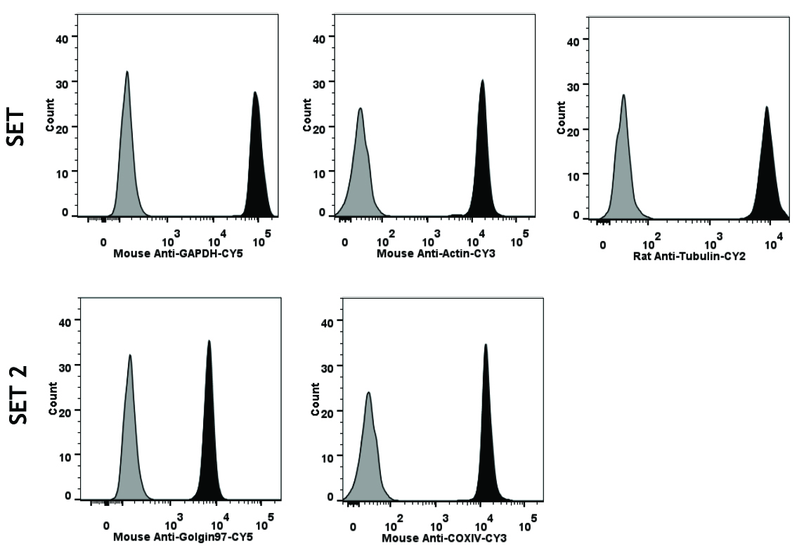
SpectraDye Antibody Labeling Kits produce antibodies comparable to commercially available fluroescent antibodies. HeLa cells were cultured in DMEM medium supplemented with FBS and Penicillin/Streptomycin. The cells were pelleted and fixed with 4% paraformaldehyde. The HeLa cells were resuspended in PBS + 0.1% Triton and SpectraDye labeled antibodies were added at 0.33µg/µL. The antibodies were incubated at ambient temperature for 30 minutes then washed two times with 1X PBS. Data was acquired with the LSRFortessa. Black filled histogram represents antibody stained cells. Black lined, gray histogram represents the unstained control.
Western Blotting: Save valuable time since secondary antibody incubation and wash steps are no longer required.
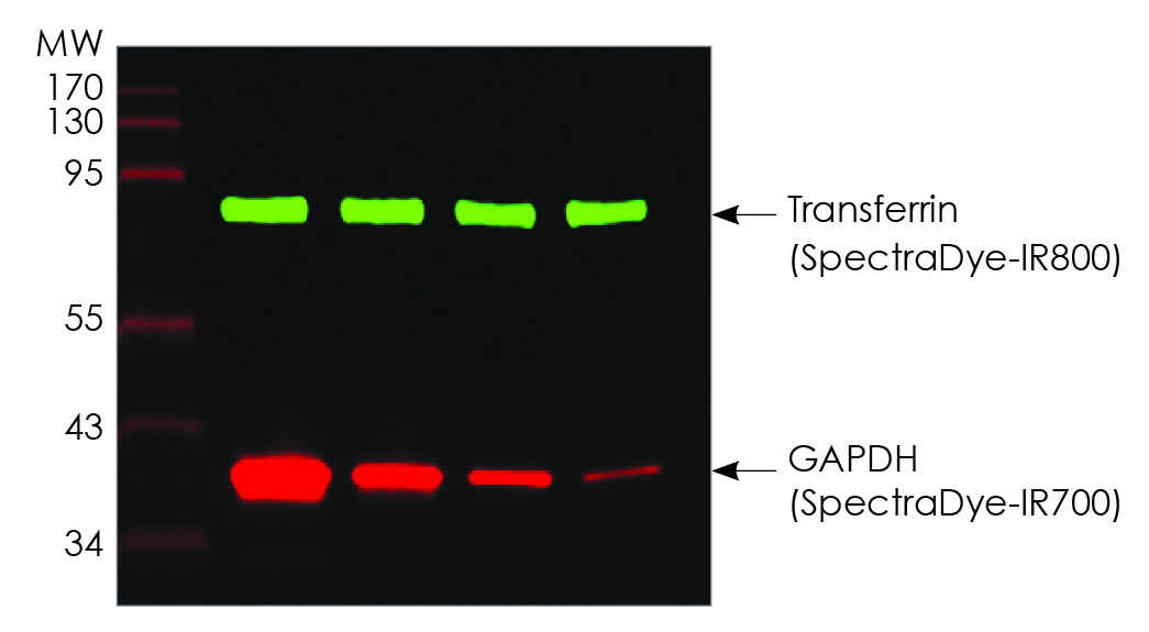
Detect two or more proteins in a single fluorescent Western blot experiment using SpectraDye-labeled primary antibodies. Transferrin (500ng/lane) and GAPDH (500, 167, 56, and 19 ng/lane) were transferred to a PVDF membrane and detected with a rabbit anti-transferrin antibody labeled with SpectraDye-IR800 and a mouse anti-GAPDH antibody labeled with SpectraDye-IR700.
SpectraDye Antikor İşaretleme Kiti BroşürüSpectraDye Antikor İşaretleme Kiti Uygulama NotuSpectraDye Antikor İşaretleme Kiti Kısa ProtokolSpectraDye Antikor İşaretleme Kiti Kolay ProtokolSpectraDye Antikor İşaretleme Kiti Dye350 MSDSSpectraDye Antikor İşaretleme Kiti Dye490 MSDSSpectraDye Antikor İşaretleme Kiti Dye547 MSDSSpectraDye Antikor İşaretleme Kiti Dye647 MSDSSpectraDye Antikor İşaretleme Kiti Dye682 MSDSSpectraDye Antikor İşaretleme Kiti Dye762 MSDSSpectraDye Antikor İşaretleme Kiti DMSO MSDSSpectraDye Antikor İşaretleme Kiti Quenching Solution MSDSSpectraDye Antikor İşaretleme Kiti Ab. Lab. Buffer MSDSSpectraDye Antikor İşaretleme Kiti Neutr. Buffer MSDS


