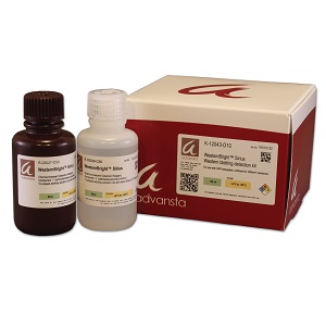
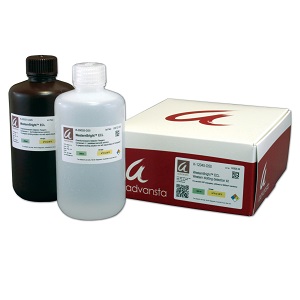
WesternBright Quantum HRP Substrat
Quantitative chemiluminescent Western blotting
• Sensitive – detect attomoles of protein per band.
• Quantitative – linear range of signal with respect to protein amount exceeds 3 orders of magnitude
• Versatile – Optimized for CCD imaging, and compatible with film detection. Chemifluorescent emissions can be detected with fluorescence imaging systems
• Low background – for high signal to noise ratio
• Long lasting signal – image blots hours after substrate incubation
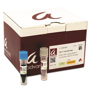
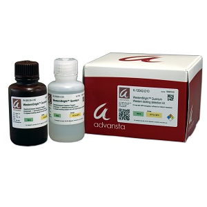
Includes:
WesternBright Quantum luminol/enhancer solution
WesternBright Quantum stabilized peroxide solution
Description
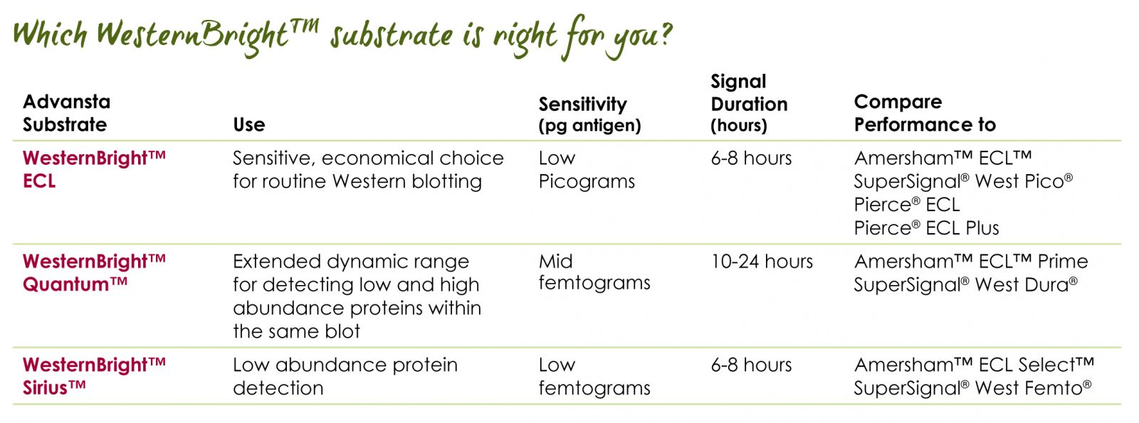
WesternBright Quantum HRP substrate
WesternBright Quantum chemiluminescent substrate sets the bar for sensitivity and quantitative ability. Specially developed for CCD imaging, WesternBright Quantum produces a strong, long-lasting signal with extremely low background, perfect for detecting low abundance proteins. Since it does not exhibit substrate depletion at high protein loads, WesternBright Quantum provides the largest dynamic range of any chemiluminescent substrate for the most quantitative chemiluminescent Western experiments.
WesternBright Quantum exhibits high sensitivity with the greatest linear range
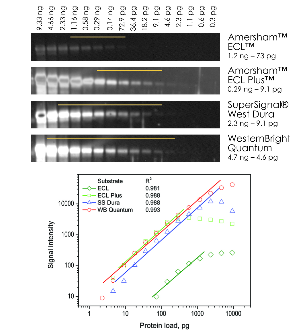
Identical Western blots containing serial dilutions of transferrin were probed with a rabbit-anti-transferrin primary antibody and a goat-anti-rabbit secondary antibody conjugated to HRP. The blots were incubated with chemiluminescent substrates as recommended by each manufacturer. All blots were imaged simultaneously for 2 min on a CCD imager and display parameters were identical across all images. Band intensities were plotted and a best fit linear regression conducted for each substrate. WesternBright Quantum shows the largest linear dynamic range out of all four substrates with the highest R2 value. Bands that fall on the linear part of the curve for each substrate are indicated in the image.
WesternBright Quantum produces the most stable chemiluminescent signal
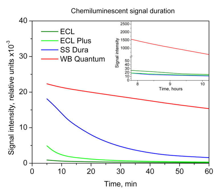
Blots detected using WesternBright Quantum or one of three other chemiluminescent substrates were re-imaged at several times over a 10 hr period. The intensitiy of one band is plotted. 60 min after substrate incubation, WesternBright Quantum retains 70% of its initial signal strength, while the competition decays to 5% or less.
Extremely low background when using WesternBright Quantum
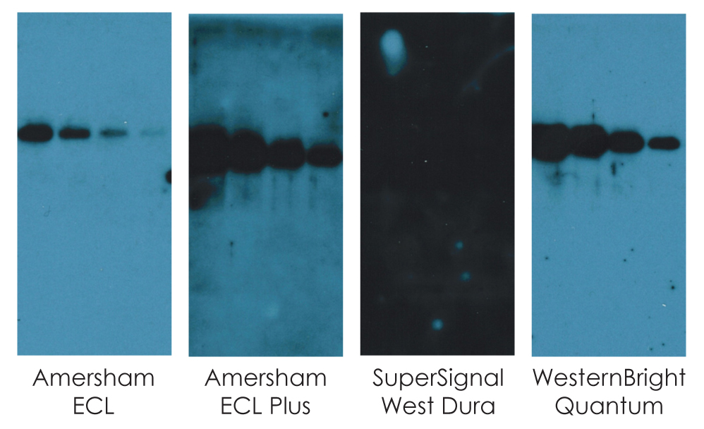
Replicate Western blots were developed using WesternBright Quantum or one of three other chemiluminescent substrates. After a simultaneous 20 min exposure to the same piece of film, WesternBright Quantum displays the best combination of sensitivity and low background signal. All display parameters were identical across all images shown in this figure.
High sensitivity for quantitative detection of low-abundance proteins
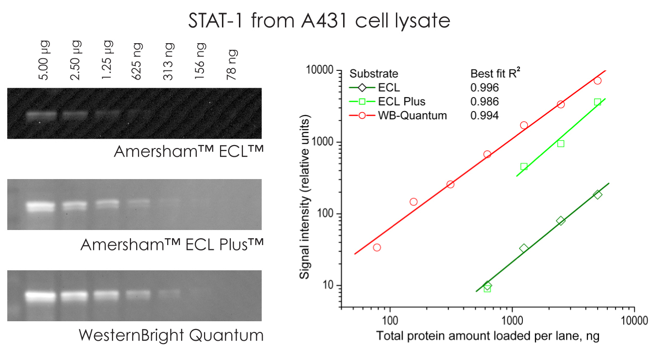
WesternBright Quantum provides the highest sensitivity and broadest linear dynamic range. Serial dilutions of A431 cell lysates were blotted and probed to detect STAT-1 protein. The blots were detected with WesternBright Quantum,or two other chemiluminescent substrates (ECL and ECL Plus, GE Healthcare). Only data points on the linear portion of the best-fit curve for each substrate shown. All display parameters were identical across all images in this figure.
No need to rush to image your Western blot
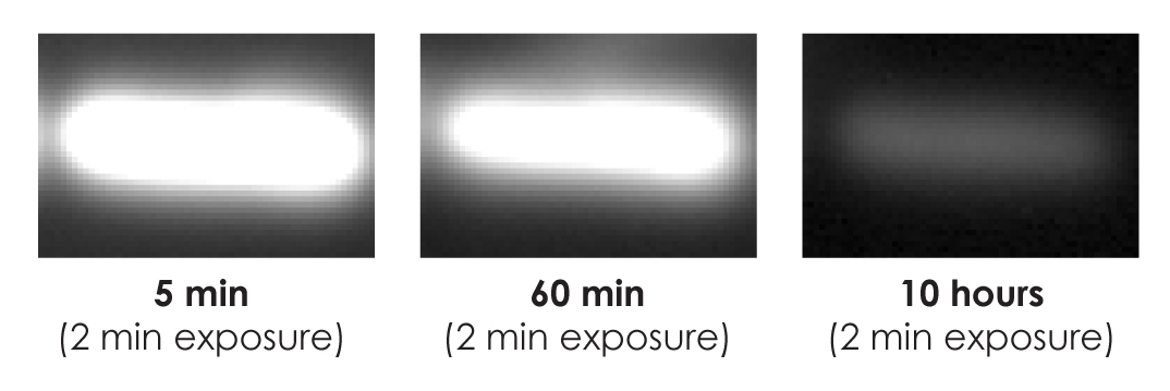
WesternBright Quantum allows blots to be imaged several hours after substrate incubation. Blots can be re-imaged to obtain the perfect exposure, without worrying about losing signal. A blot was imaged with 2 min exposures at 5 min, 60 min, and 10 hours after substrate incubation. The same band is clearly seen in each image, even after 10 hours.
Superior sensitivity with film
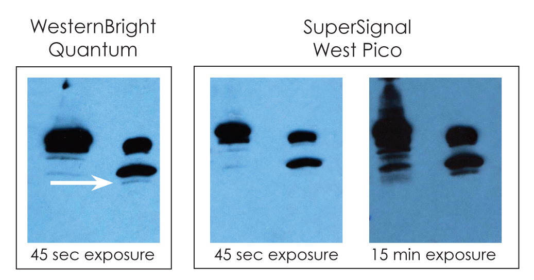
WesternBright Quantum provides high-performance film detection.WesternBright Quantum or SuperSignal® West Pico (Thermo Scientific) were used to detect duplicate Western blots. A band (arrow) is detected by WesternBright Quantum in a brief exposure, while a much longer exposure is needed to detect the same band with SuperSignal West Pico. All display parameters were identical across all images in this figure


