AdvanStain Scarlet
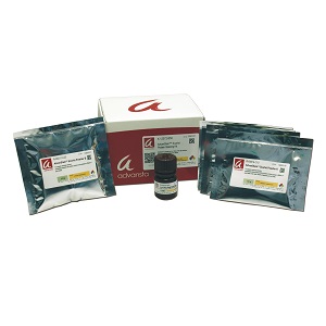
Fluorescent protein stain for gels and blots
• Sensitive – detect less than a nanogram of protein per spot
• Convenient – simple 3-hour protocol
• Flexible – stain gels or membranes
• Reversible – destain for downstream Western blotting or mass spectrometry
• No speckling – clean background for better data
• Safe – biodegradable and contains no heavy metals for increased safety and simple disposal
• Compatible – image with any laser or CCD imaging system. Compatible excitation wavelengths include green (543, 532 nm), blue (488 nm), violet (405 nm) and UV (302, 365 nm). Maximum emission wavelength is 610 nm
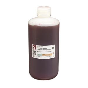
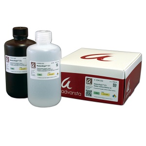
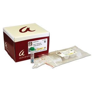
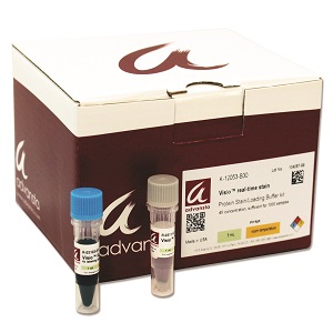
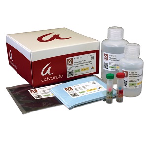
*Includes: AdvanStain Scarlet Dye Concentrate; Powder A for preparation of Fix Solution; Powder B for preparation of Stain Buffer.
Description
AdvanStain Scarlet is a fluorescent protein stain for gels and blots that allows sensitive and quantitative visualization of protein bands. With a quick and easy protocol, gels can be stained in less than 3 hours, and imaged on a wide variety of imaging systems. Gels and blots stained with AdvanStain Scarlet can be imaged on any fluorescence imaging system, including laser- and CCD-based systems, with a detection limit of less than 1 ng of protein per band or spot. Fully compatible with downstream Western blotting or mass spectrometry, AdvanStain Scarlet is non-toxic and biodegradable, for safe and simple disposal.
AdvanStain Scarlet shows sensitivity and linearity
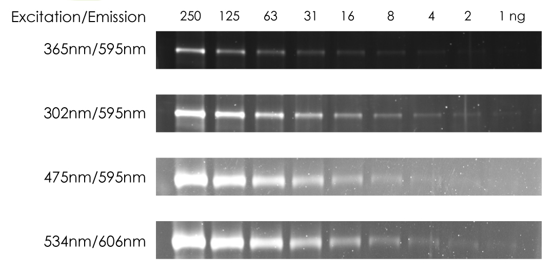
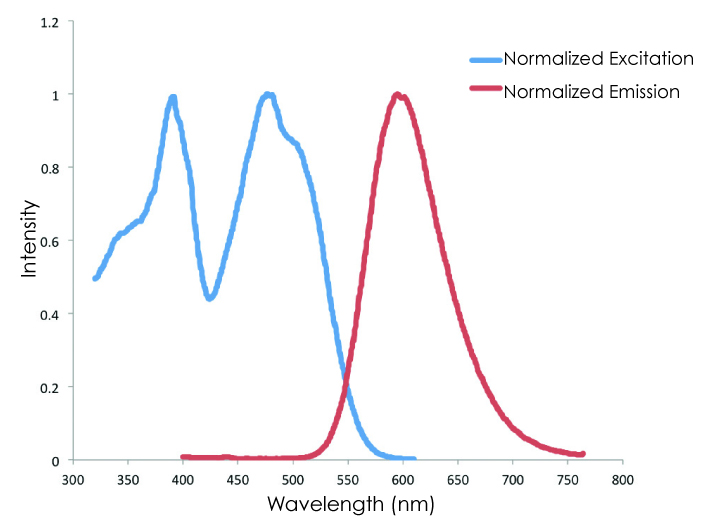
Figure 1. AdvanStain Scarlet can be imaged under multiple imaging conditions, with sensitivity of 1 ng or less per band. The optimal excitation and emission wavelengths of AdvanStain Scarlet are compatible with multiple imaging conditions, and gels stained with AdvanStain Scarlet can be imaged using UV (305 or 365 nm), blue, or green-light excitation, using a transilluminator, CCD- or laser-based imaging system.
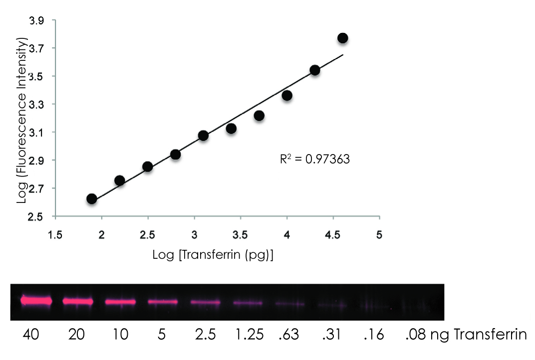
Figure 2. AdvanStain Scarlet signal is linear for more than 2 orders of magnitude. Serial dilutions of transferrin protein were analyzed by SDS-PAGE and the gel stained with AdvanStain Scarlet.
AdvanStain Scarlet outperforms other protein stains
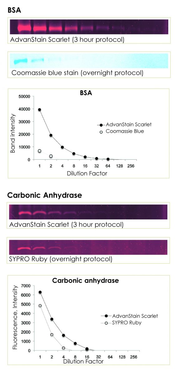
Figure 3. AdvanStain Scarlet outperforms both Coomassie and Sypro Ruby. The gels were stained with AdvanStain Scarlet, SYPRO Ruby, or Coomassie blue stain according to manufacturer’s instructions, and then imaged on a CCD imager.
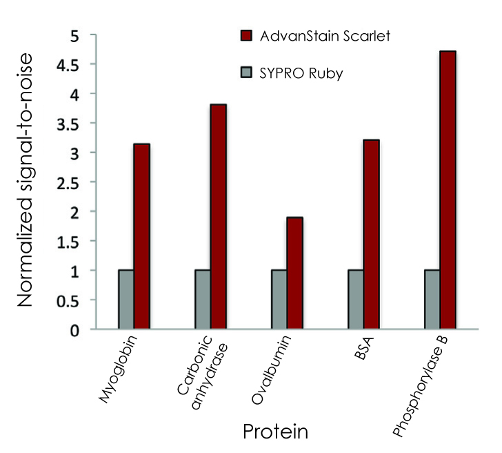
Figure 4. AdvanStain Scarlet provides a higher signal-to-noise ration than SYPRO Ruby. Serial dilutions of several proteins were separated on duplicate 1D gels, then stained with either AdvanStain Scarlet or SYPRO Ruby according to each manufacturer’s instructions. For each protein, signal-to-noise values were normalized to the value obtained for the protein on the SYPRO Ruby-stained gel. AdvanStain Scarlet provided a signal-to-noise ratio at least 2-times greater than SYPRO Ruby for each protein. The lower background observed with AdvanStain is responsible for the higher signal-to-noise ration, increasing confidence in quantitiation of low-abundance bands and spots.


