İnkübasyon Kutuları
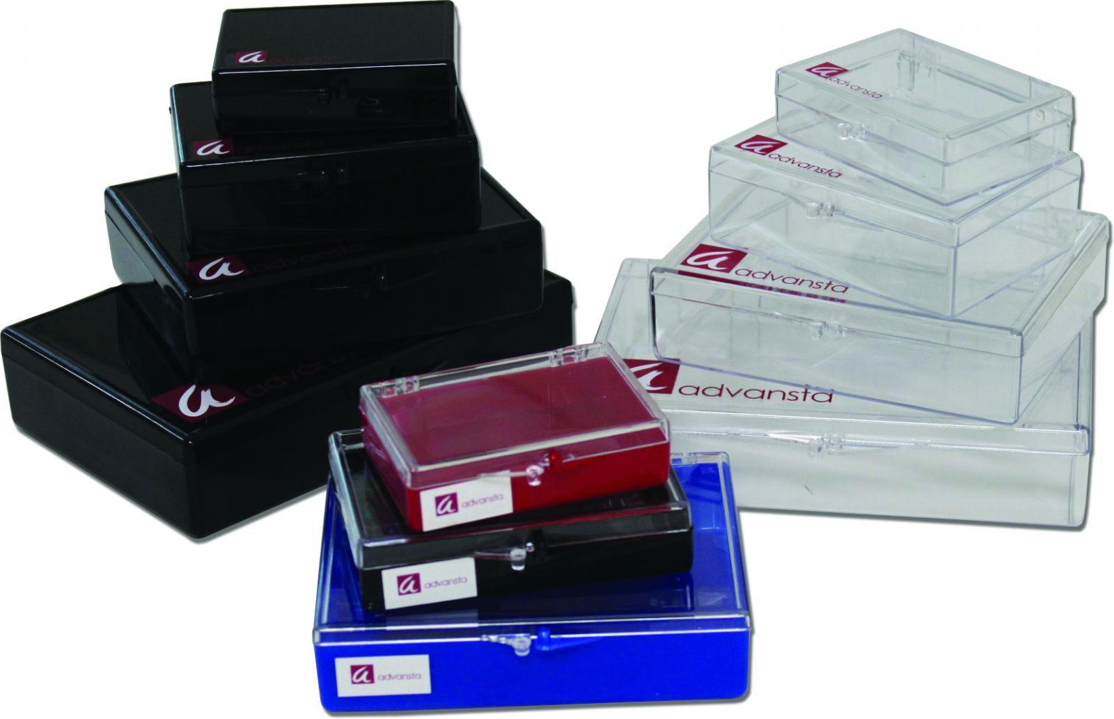
Convenient containers for staining and washing gels and membranes
• Smooth interior – protects membranes and gels from scratches
• Convenient – multiple sizes (5×7, 6×9, 9×11,10.5×15.5 cm) and designs available for different gels and blots
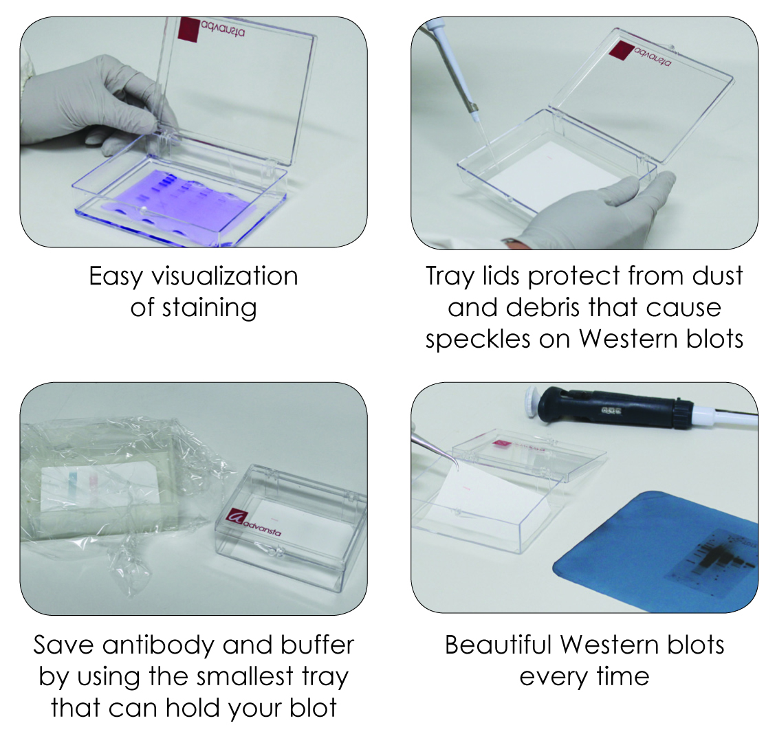
• Attached lid – to protect experiments from dust or debris that can cause speckles on blot images
• Save – minimize antibody and buffer usage by using appropriately sized containers
• Multi-chamber trays – process 6 mini-blots simultaneously, each chamber requires as little as 3 mL of solution
Description
Incubation trays
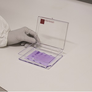
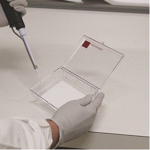
These lidded incubation trays have a perfectly smooth interior surface, making them ideal for staining and washing electrophoresis gels and membranes. Trays are manufactured to prevent adsorption of antibodies.
Choose from three designs, all available in a range of sizes:
• Opaque, for light-sensitive applications
• Transparent, for easy monitoring of colorimetric staining
• Traditional, for everyday blot washes and incubations
The three available designs are all available in a range of sizes. Opaque containers are ideal for light-sensitive applications; transparent containers are ideal for easy monitoring of colorimetric staining; traditional containers are convenient for typical blot washes and incubations.

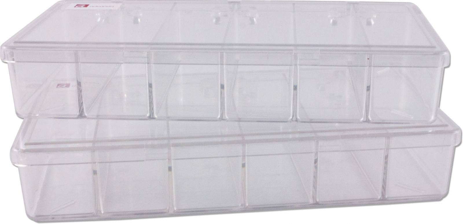
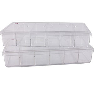
Multi-chamber incubation trays
These specially designed incubation trays are ideal for staining and washing electrophoresis gels and membranes that have been partitioned. Each Multi-chamber incubation tray is partitioned into six chambers that measure 2.7 x 8.1 cm.

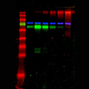
Western blot membranes processed in Multi-chamber incubation trays are comparable to standard incubation trays. HeLa cell lysate was separated by SDS-PAGE and transferred to a PVDF membrane. An intact membrane was processed side-by-side a membrane that was partitioned and incubated in three chambers of a multi-chamber incubation tray. The membranes were blocked with AdvanBlock™- Chemi before incubation with a rabbit anti-human STAT-1 (Millipore #06-501) primary antibody. Signal was detected with WesternBright® ECL substrate.


